Removing the Friction in documenting Ocular Structures.
Discover how eye care practices can improve ocular structure documentation with digital drawing tools, grading scales, image uploads, cup-to-disc ratio capture, and customizable pre-filled clinical findings.
In busy clinics, ocular structure documentation can slow everything down: exam room flow, report quality, and handoffs between clinicians. Most ophthalmology and optometry teams know the challenge well: drawing findings quickly, documenting consistently, and still keeping records clinically useful.
Industry literature reflects this pain. The American Academy of Ophthalmology has highlighted that ophthalmology is visually intensive and needs EHR workflows that support image-based and drawing-heavy documentation, not text-only forms (AAO special requirements paper). Studies have also shown that documentation methods can become slower or harder to use when they do not fit how eye exams are actually recorded (PMC study, JAMA Ophthalmology / PMC).
ASIRA has designed the ocular structure workflow specifically for this reality.
The biggest documentation pain points in eye care
Clinicians and administrators often face five recurring issues in ocular structure reporting:
- Drawing friction: generic charting tools make it hard to quickly mark lesions, tears, vascular changes, or staining patterns.
- Inconsistent grading: without visual grading references, cataract and lens findings vary between providers.
- Disconnected imaging: slit lamp or fundus photos are often stored separately from exam notes.
- Critical glaucoma data lost in text: cup-to-disc ratio capture is inconsistent or buried in free text.
- Rigid dropdowns: teams need standardization, but also the ability to add clinic-specific findings.
How ASIRA solves ocular structure documentation
Built-in drawing tools for anterior and posterior segment documentation
Drawing tool for Anterior Segment Evaluation
ASIRA provides dedicated ocular diagram workflows with practical drawing controls: pen, eraser, annotation text, arrows, outline markers, and condition-specific visual stamps. This means clinicians can document what they see in real time, visually, without leaving the exam workflow.
For anterior segment evaluation and posterior segment evaluation, diagram markup is not an afterthought. It is part of the core charting experience, which supports faster and more readable clinical documentation.
LOCS III-friendly crystalline lens grading support
ASIRA includes a LOCS III-friendly grading interface for crystalline lens evaluation with structured choices for nuclear, cortical, and posterior categories. That makes cataract grading more consistent across clinicians and easier to track longitudinally.
When grading systems are embedded directly in workflow, practices reduce variability and improve report clarity for referrals, surgery planning, and follow-up.

Upload existing patient photos directly into the exam workflow
Clinicians can upload existing patient images (including slit lamp images) into ocular structure evaluation views. This helps practices keep visual evidence linked to exam documentation instead of scattered across systems or devices. For administrators, this improves record completeness and supports better audit readiness and continuity of care.
Structured cup-to-disc ratio capture for glaucoma workflows
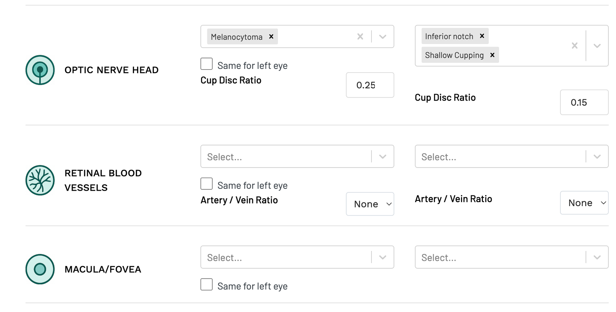
ASIRA includes dedicated Cup/Disc Ratio fields for right and left eye under optic nerve head documentation. Instead of relying on narrative-only entries, teams can now record C/D ratio in a structured, reportable format that supports glaucoma monitoring over time.
This is especially useful for trend tracking, inter-clinician consistency, and cleaner downstream reporting.
Large pre-filled condition lists plus custom additions
ASIRA starts with an extensive list of ocular findings across key structures (anterior segment, posterior segment, crystalline lens) and lets your team add new observations when needed. So you get both standardization and flexibility: fast selection for common findings, plus customization for real-world practice variation.
Why this matters for clinics and hospitals
Better ocular structure documentation is not just a clinician convenience. It impacts:
- visit efficiency,
- chart quality,
- referral communication,
- medico-legal clarity,
- and operational consistency across providers.
ASIRA's ocular structure workflow is built for how eye care teams actually examine, draw, grade, and document.
If your practice is looking to improve ophthalmology documentation quality while reducing charting friction, this is exactly where modern workflow design makes a measurable difference.
To Book a Demo or to learn more, contact ASIRA at contact@asira.health or via WhatsApp: +919152391194. Visit www.asira.health to learn more about how ASIRA can help your eye care business grow.


Comments ()