Artificial Intelligence in Diabetic Retinopathy Screening: A Comprehensive Overview
Diabetic retinopathy (DR) is a leading cause of blindness among working-age adults globally. Early detection and timely intervention are crucial in preventing vision loss associated with DR. Traditional screening methods often involve manual assessment of retinal images by trained specialists, which can be time-consuming and subject to human error. The advent of Artificial Intelligence (AI) has introduced new possibilities in the early detection and management of DR. This article explores the role of AI in DR screening, focusing on the FDA-approved IDx-DR system, its clinical applications, and future prospects.
Understanding Diabetic Retinopathy
Diabetic retinopathy is a microvascular complication of diabetes mellitus, characterized by damage to the blood vessels in the retina. It progresses through four stages: mild nonproliferative, moderate nonproliferative, severe nonproliferative, and proliferative diabetic retinopathy. Early stages may be asymptomatic, making regular screening essential for individuals with diabetes. Without timely intervention, DR can lead to significant vision impairment and blindness.
The Role of Artificial Intelligence in DR Screening
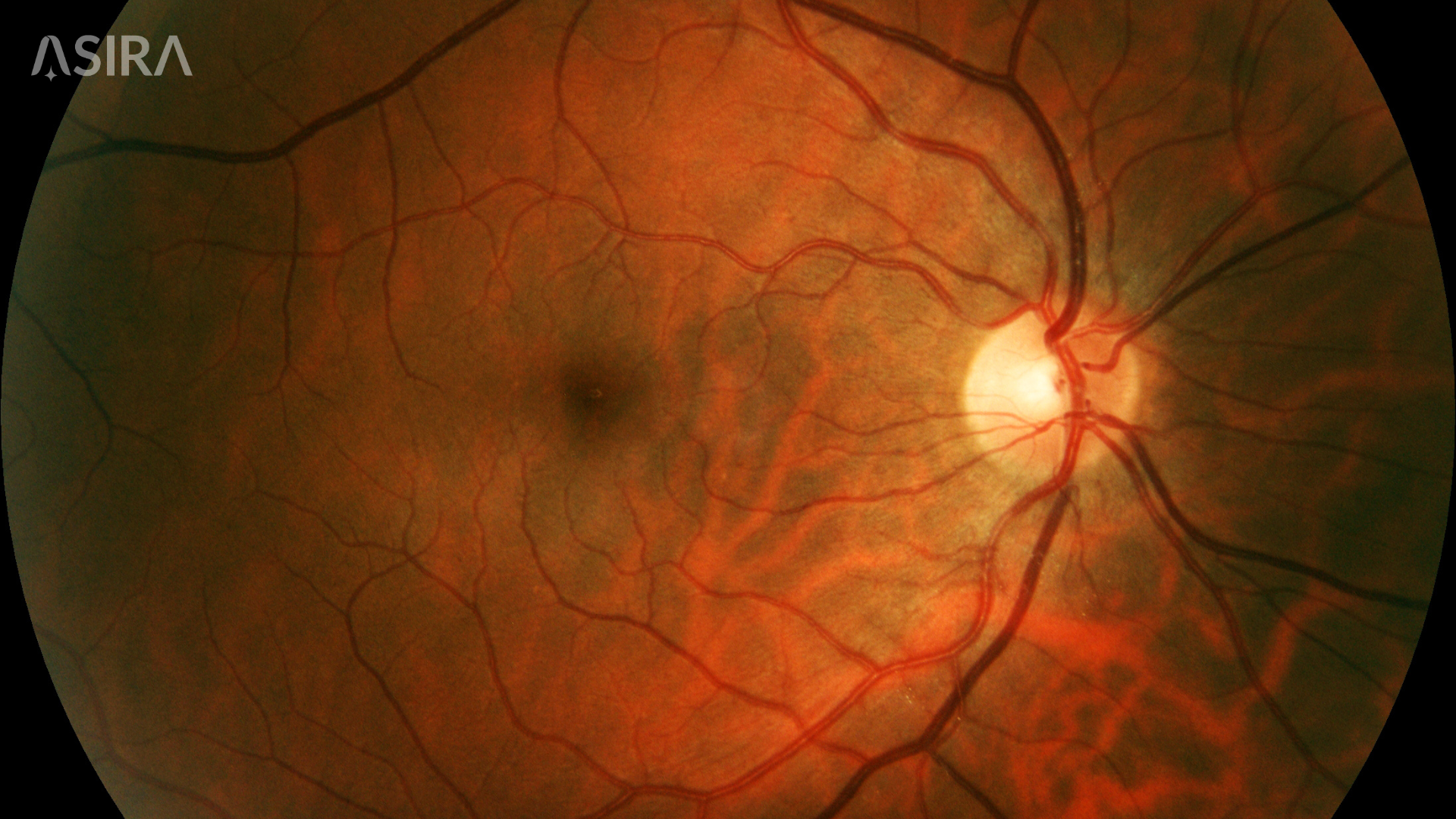
Artificial Intelligence, particularly deep learning algorithms, has shown promise in automating the detection of DR. These AI systems analyze retinal images to identify signs of DR, such as microaneurysms, hemorrhages, and exudates. By training on large datasets of labeled retinal images, AI models can learn to recognize patterns indicative of DR.
The IDx-DR system, developed by Digital Diagnostics, is an AI-based diagnostic tool designed to detect more than mild diabetic retinopathy. It was the first autonomous AI system authorized by the U.S. Food and Drug Administration (FDA) for use in primary care settings. The system analyzes retinal images captured using a fundus camera and provides a result within minutes, indicating whether the patient has referable DR.
Clinical Validation of IDx-DR
The diagnostic accuracy of IDx-DR has been evaluated in several clinical studies. A pivotal trial demonstrated that IDx-DR had a sensitivity of 87.2% and a specificity of 90.7% in detecting more than mild DR when compared with grading of wide-angle stereoscopic fundus photographs by the University of Wisconsin Fundus Photography Reading Center. In comparison, the sensitivity and specificity of a dilated ophthalmic exam by board-certified ophthalmologists were 34% and 100%, respectively, when compared with a similar, outcome-based reference standard of stereoscopic mydriatic fundus photos graded by a reading center. This indicates that IDx-DR was more sensitive than ophthalmologists in identifying early DR.
Further studies have supported these findings, showing that IDx-DR maintains high diagnostic performance across diverse populations and clinical settings. For instance, a systematic review and meta-analysis involving over 13,000 participants reported pooled sensitivity and specificity values of 0.95 and 0.91, respectively, for IDx-DR in detecting DR.
Implementation in Clinical Practice
Integrating AI systems like IDx-DR into clinical practice requires careful consideration of various factors. Training healthcare providers to operate the system and interpret its results is essential. While IDx-DR provides an assessment of retinal images, the final diagnosis and treatment decisions should remain with qualified clinicians. AI should be viewed as a supportive tool that augments human expertise, not as a replacement.
Moreover, establishing protocols for the use of AI in DR screening is crucial. This includes determining patient eligibility, ensuring quality control of retinal images, and addressing any technical issues that may arise. Regular calibration and maintenance of the AI system are also necessary to maintain its accuracy and reliability.
Benefits of AI in DR Screening
The adoption of AI in DR screening offers several advantages:
- Increased Access: AI systems can be deployed in primary care settings, extending screening to underserved populations and areas with limited access to ophthalmologists.
- Efficiency: Automated analysis of retinal images reduces the time required for screening, allowing for higher throughput and timely interventions.
- Consistency: AI algorithms provide consistent assessments, minimizing variability associated with human interpretation.
- Early Detection: AI can identify subtle signs of DR that may be overlooked by human examiners, facilitating earlier intervention and better patient outcomes.
Challenges and Considerations
Despite its potential, the implementation of AI in DR screening presents several challenges:
- Data Quality: The accuracy of AI systems depends on the quality of the input data. Poor-quality retinal images can lead to incorrect assessments.
- Integration with Existing Systems: Integrating AI tools into existing electronic health record systems can be complex and may require significant resources.
- Regulatory and Legal Issues: The use of AI in medical diagnostics raises questions about liability and accountability. Clear guidelines and regulations are needed to address these concerns.
- Ethical Considerations: Ensuring that AI systems are free from bias and equitable across different populations is paramount. Ongoing monitoring and validation are necessary to maintain fairness.
Future Directions
The future of AI in DR screening is promising. Advancements in deep learning techniques and the availability of larger, more diverse datasets will likely improve the accuracy and generalizability of AI models. Additionally, the integration of AI with other diagnostic tools, such as optical coherence tomography (OCT), could provide a more comprehensive assessment of retinal health.
Furthermore, AI has the potential to play a role in predicting the progression of DR and personalizing treatment plans. By analyzing longitudinal data, AI systems could identify patients at high risk of disease progression, enabling proactive management strategies.
Conclusion
Artificial Intelligence is transforming the landscape of diabetic retinopathy screening. Systems like IDx-DR have demonstrated high diagnostic accuracy and offer the potential to increase access to screening, particularly in underserved areas. However, successful implementation requires careful planning, ongoing validation, and collaboration between AI developers and healthcare providers. As technology continues to evolve, AI is poised to play an increasingly integral role in the early detection and management of diabetic retinopathy, ultimately improving patient outcomes and reducing the burden of vision loss associated with diabetes.
References
Abràmoff MD, et al. Pivotal trial of an autonomous AI-based diagnostic system. JAMA Ophthalmol. 2018;136(2):118-126. doi:10.1001/jamaophthalmol.2017.5565.
Khan Z, et al. Diagnostic accuracy of IDX-DR for detecting diabetic retinopathy: A systematic review and meta-analysis. Am J Ophthalmol. 2025;160(1):1-10. doi:10.1016/j.ajo.2025.02.022.
Channa R, et al. Autonomous artificial intelligence in diabetic retinopathy screening. JAMA Ophthalmol. 2020;138(3):268-274. doi:10.1001/jamaophthalmol.2019.7039.
Rajesh AE, et al. Artificial intelligence and diabetic retinopathy: AI framework, prospective studies, head-to-head validation, and cost-effectiveness. Diabetes Care. 2023;46(10):1728-1739. doi:10.2337/dci23-0029.
Ursin F, et al. Diagnosing diabetic retinopathy with artificial intelligence. Ophthalmology. 2021;128(3):383-391. doi:10.1016/j.ophtha.2020.08.022.


Comments ()